Where Science Encounters Art
The biological world is not only intellectually captivating but also visually stunning. The techniques and tools we employ in our research enable us to gain profound insights and also observe the beauty that unfolds from our experimental specimens. Here we showcase some of the images from our research, most of them were captured using confocal microscopy. Some of the images have been recognized with awards.

The beauty lies in the folds
Mouse cerebellum labelled with mRNA encoding key synaptic proteins teneurin and latrophilin (yellow and red). Cell nuclei is shown in blue.

Intricate yet structured
Sagittal section of mouse brain labelled with mRNA encoding synaptic proteins teneurin and latrophilin een, red, purple). Read associated publication here

Cellular cosmos
mRNA belong to three synaptic organizers from same gene family labelled in green, red and purple in retrosplenial area of mouse brain

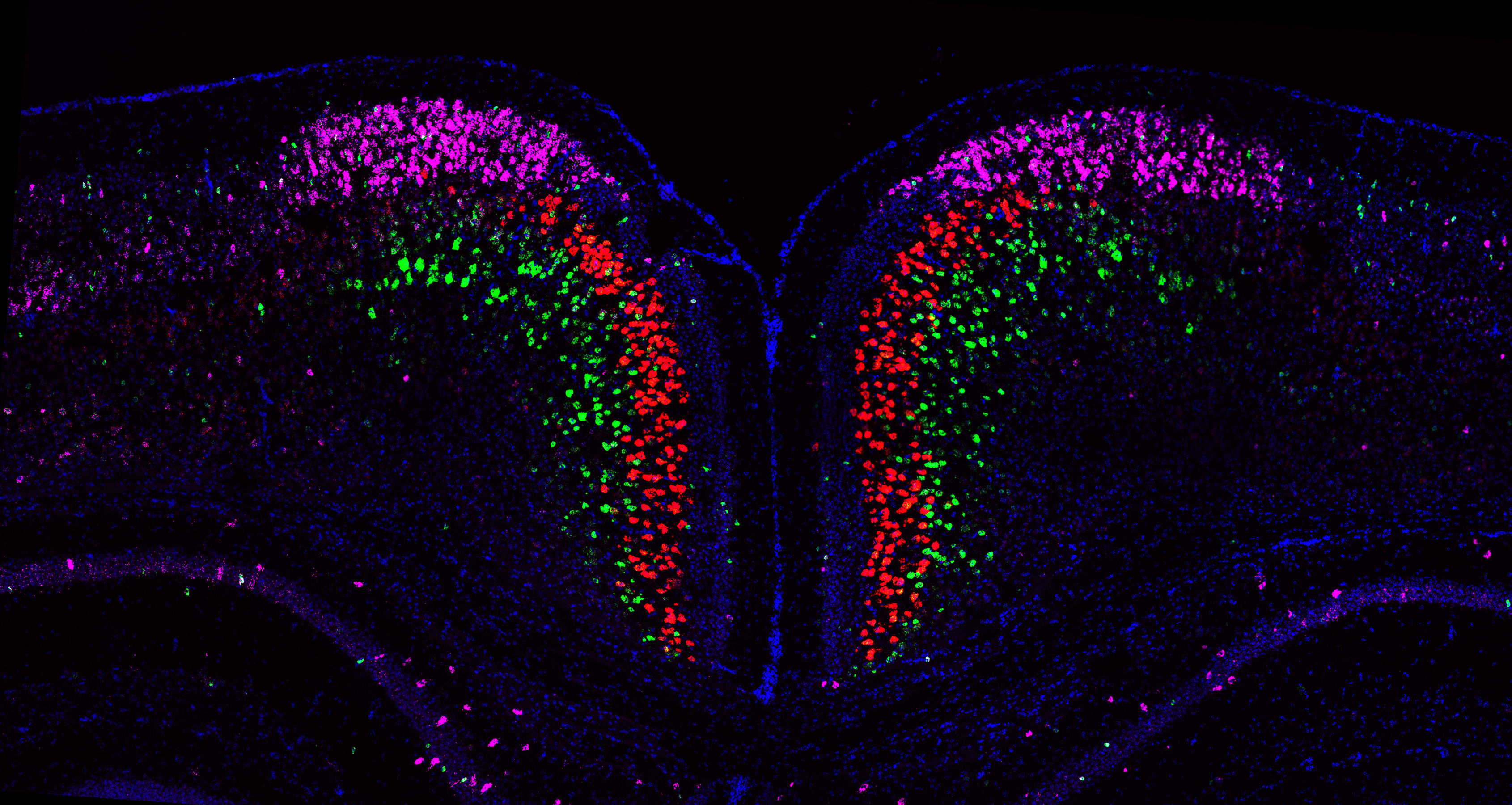
Mirroring Molecules
mRNA belong to three synaptic molecules from same gene family labelled in green, red and purple in olfactory bulb of mouse brain

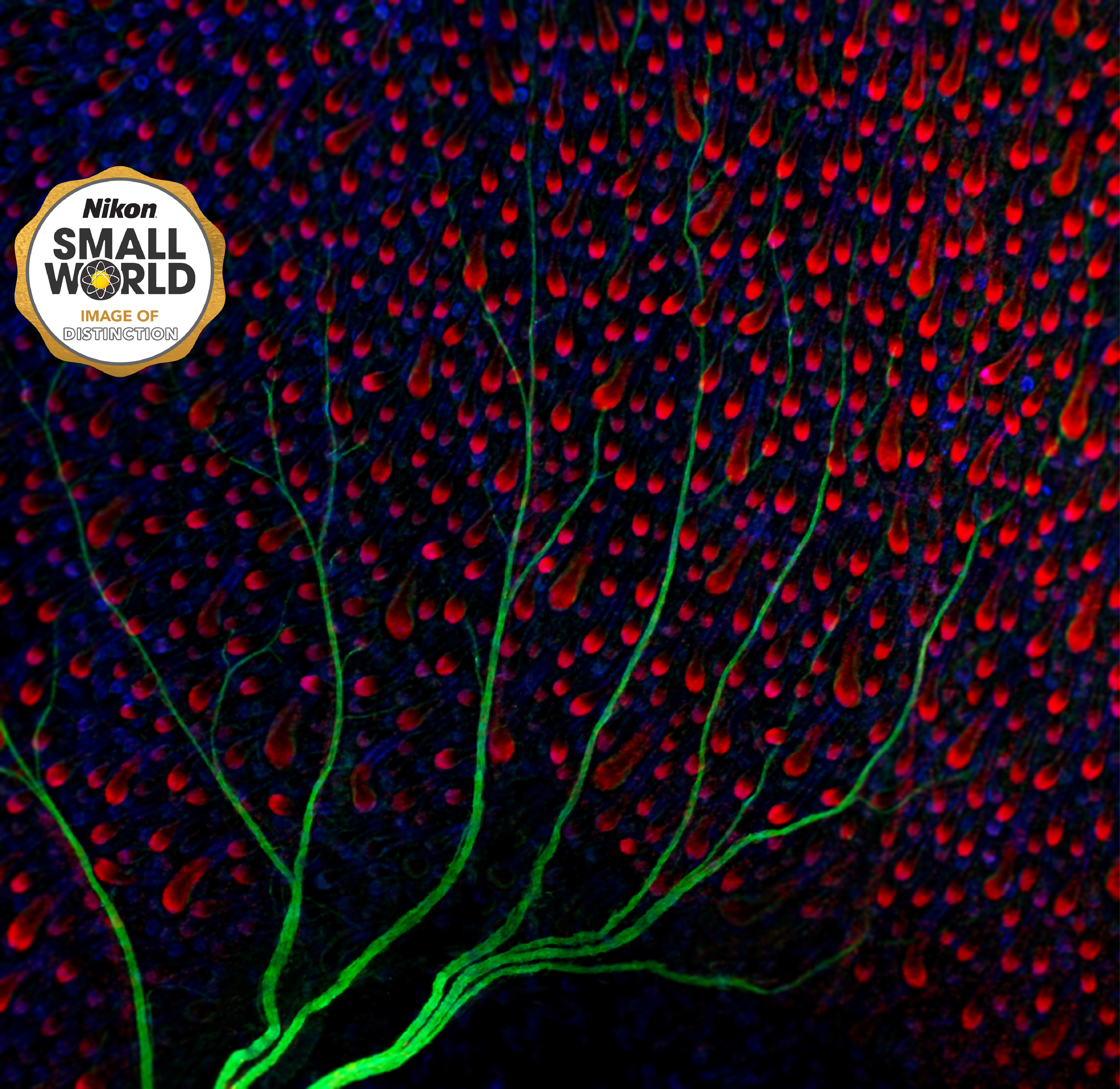
Skin Tree
Nerve (in green) under the skin of mouse (hair follicles are shown in red and blue).
Image of Distinction

Inner artwork of a mouse eyelid
Unopened eye lid of a newborn mice labelled with markers for epidermal sheet and hairfollicles.
Finalist - Micro category

Star-studded skin
Melanocytes (in green) scattered through the epidermis and the hair follicle (in red).
1st Prize - British Society for Cell Biology Image Competition 2015

Deep rooted goose bumps
The arrector pili muscle aka goose bump muscle fibres (in green) attached to hair follicles (purple cells) of mouse skin.

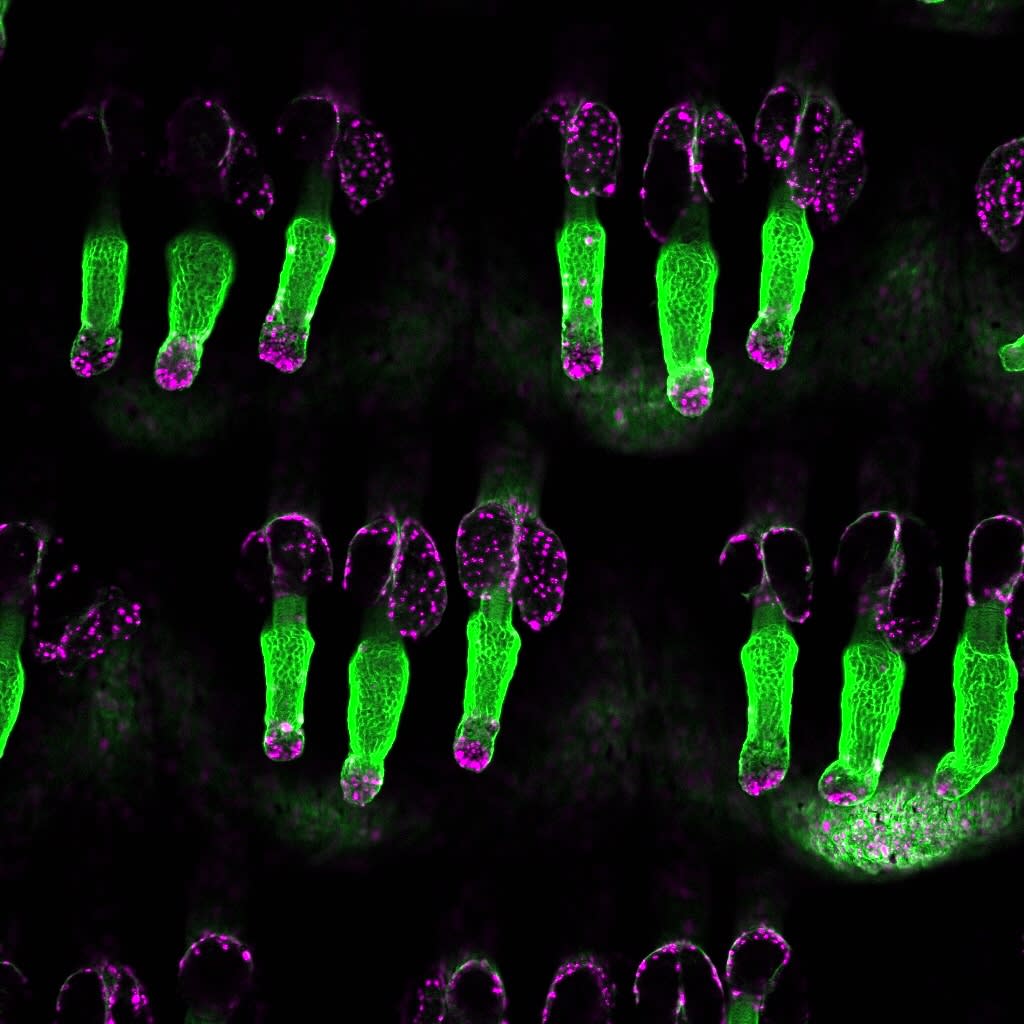
Glowing in the dark
Hair follicles (in green) and rapidly dividing stem cells are labelled in purple.

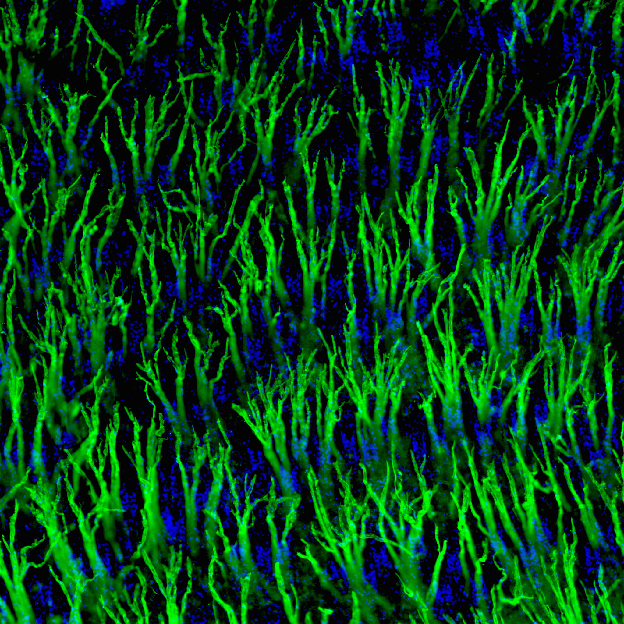
Looking for connection
Top view of arrector pili muscle aka goose bump muscle fibres (in green) attached to hair follicles (blue cells) of mouse skin.


